The
normal ocular surface is covered by epithelial cells which can be1
damaged by certain systemic inflammatory diseases,1 primary ocular
diseases, and trauma resulting in the breakdown of ocular surface.2 If
the normal epithelialization process fails ocular defect becomes chronic.
Chronic inflammation leads to neovasculari-zation, corneal scarring,
opacification, corneal thinning, and possible corneal perforation.
Traditional
treatments for ocular surface disorders include correcting underlying
pathology, suppressing inflammation and promoting healing process. Currently,
artificial tears, lubricants, fibronectins,3,4 growth factors,5
and substance P6 are used. However, if defect persists and stromal
thinning develops, more invasive surgical options like tissue adhesive7,
bandage contact lens,8 conjunctival flap9, and
tarsorrhaphy can be performed10. But these treatments have their own
complications. In this background amniotic membrane can be considered as an
option for treating the ocular surface defects3,4.
In
1910, Davis reported the use of fetal membrane in skin transplantation for the
first time11. Amniotic membrane transplantation in
ophthalmology was reported by De Roth in 1914 who achieved partial success in
treatment of conjunctival epithelial defects12. There was
very little information available in ophthalmic literature until the study by
Kim and Tseng in 1995 who used amniotic membrane transplantation for ocular
surface reconstruction of severely damaged cornea in rabbit model. Since that
experimental study, amniotic membrane transplant-tation has been used for
persistent corneal epithelial defects, neurotrophic corneal ulcers,
conjunctival surface reconstruction, bullous keratopathy, chemical or thermal
burns and in patients of Steven-Johnson syndrome13-15.
Ocular surface disorders are a
common problem and current management is not satisfactory. Amniotic membrane
transplantation has shown better results in treating these disorders. In
MATERIAL AND METHODS
This Case series was conducted
at
Preparation of Amniotic membrane:
Amniotic membrane was obtained
from prospective donors undergoing Caesarean section, who were negative for
communicable diseases including HIV, hepatitis and syphilis. Different
protocols exist for the processing and storage. We used
protocol described by Kim et al16. According
to which placenta is cleaned and stored with balanced salt solution containing
a cocktail of antibiotics (Table 1) under sterile conditions.
Surgical Techniques
I.
Inlay or graft technique: When Amniotic membrane is tailored to the
size of the defect, is meant to act as a scaffold for the epithelial cells and
which then merges with the host tissue, it is referred to as a graft.17
Amniotic membrane was secured with its basement membrane or epithelial side up
to allow migration of the surrounding epithelial cells on the membrane (Fig. 1).
II. Overlay or patch technique: When
the Amniotic membrane is used akin to a biological contact lens in order to
protect the healing surface defect beneath; it is referred to as a patch18.
A patch also reduces inflammation by its barrier effect against the chemical
mediators from the tear film. When used as patch the membrane is secured with
its epithelial side up and it either falls off or is removed.
III. Filling-in or layered technique: In
this technique the entire depth of an ulcer crater is filled with small pieces
of AM trimmed to the size of the defect. A larger graft is sutured
to the edges of the defect in an inlay fashion and an additional patch may help
in preserving the deeper layers for a longer duration19.
Preoperative evaluation was applied to all patients with special attention
given to patient’s symptoms with respect to pain and photophobia, best
corrected visual acuity. Follow up was done at first post operative day,
1st week, 2nd week and 1 month for best corrected visual
acuity, ocular symptoms (pain and photophobia) and complications. The data was analyzed by SPSS version
10.00, the variables of outcome measures (pain, photophobia, best corrected
visual acuity, graft uptake) was presented as proportions and ratios. The
variables of outcome were compared with some of variables of demography. Since
this study was a quasi experimental, no test of significance was necessary.
RESULTS
Of the
30 patients of different ocular surface disorders 18 were males (60%) and 12
were females (40 %). Ocular surface disorders of various types were included in
this study, most was the bullous keratopathy 8 (26.7%) followed by Mooren’s
ulcer 5 (16.7%), Shabbir syndrome 5 (16.7%), impending perforations 4(13.3%),
Chemical injury 3 (10.0%), Steven Johnson syndrome 3 (10%) and 2 (6.7%) cases
of neurotrophic ulcer.
The ocular surface defects was present in both eyes of 9 (30.0%)
cases. 13 (43.3%) cases had these defects in right eye, while 8 (26.7%) cases left eye was involved out of total 30
cases.
Ocular
pain was one of the most important variable of study. It was recorded on the
pain scale from grade 0 – 4 as described by the patient. Three (10.0%) patients
did not complain any pain (Grade 0). Six (20.0%) cases had mild pain (grade
1).Seven (23.3%) cases were having moderate pain (Grade 2). Thirteen (43.3%)
patients described severe pain. One (3.3%) case was having maximum pain
imaginable (Fig.2). After one month of amniotic membrane transplantation, most
of the patients 25 (83.3%) were having no pain (Grade 0). Only 2 (6.7%) and 3
(10.0%) patients described mild (Grade 1) and moderate (Grade 2) pain. No
patient described grade 3 and 4 level of pain (Fig. 3).
Twenty seven (90%) of the patients were photophobic, only 3
(10.0%) out of 30 did not complain of photophobia. A remarkable improvement was
noted in this regard. At one month after surgery, 26 (86.7%) patients did not
complain of photophobia and only 4 (13.3%) cases were still complaining of it.
There
was a little improvement of best corrected visual acuity noted, after 1month of
surgery 4 (13.3%) had best corrected visual acuity 6/12, while 1 (3.3%) case
had 6/18 and 2 (6.7%) patients were having 6/24. Majority of the cases 23 (67%)
were still having best corrected visual acuity 6/60 or less.
Table 1: Contents and concentrations of
antibiotics solution
|
Antimicrobial Agent |
Dose |
|
Penicillin |
50 mg/ml |
|
Streptomycin |
50 µg/ml |
|
Neomycin |
100 mg/ml |
|
Amphotericin B |
2.5 mg/ml |
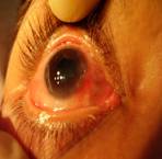
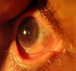
A B
Fig.1: Inlay technique used on Mooren’s ulcer
A.
Pre operative B. Post operative
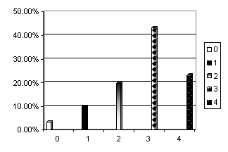
Fig. 2: Pre Operative Pain
Grade

Fig. 3: Post Operative pain
grade
DISCUSSION
Ocular surface disorders are a common problem that presents
not only with decrease of vision but also pain and photophobia. Unfortunately,
its currently medical or surgical treatment has not shown satisfactory results
so far. Amniotic membrane that had been used for other purposes like biological
dressing to cover the open wounds and skin transplantation, have also shown
good results in ocular surface defects healing and thus relieving the symptoms
of ocular irritation.
Human
amniotic membrane is derived from the fetal membranes and is loosely attached
to the chorion. 20 It is composed of three layers: a single
epithelial layer, thick basement membrane, and a vascular stroma. Human
amniotic membrane has been shown to contain collagen types III and V. It also
contains collagen types IV and VII similar to corneal epithelial basement
membrane as well as fibronectin and laminin21. Additionally, it
contains fibroblast and other growth factors. Amnion prevents inflammatory
cell infiltration and reduces apoptosis in
keratocytes after transplantation onto the corneal surface22. Due to all these properties
amniotic membrane transplantation is found to be an important tool for
reconstruction of ocular surface disorders.
Reduction
in symptoms of ocular irritation that includes pain and photophobia was 90 % in
our study which is comparable to the other studies23. Increased
comfort level, improved the quality of life of the patients. There was no
remarkable improvement in best corrected visual acuity observed in our study.
The final visual acuity less than 6/60 was recorded in 67 % of cases in our
study which was quite similar to study by Prabhasawat P, Tesavibul N who also
observed the similar ratio in their study23. However increased
comfort level improved the quality of life of these patients and visual acuity
was not the issue in these patients.
Failure
was noted in 3 (10%) cases in our study. This was due to graft necrosis, active
infection and intractable corneal perforation. This failure points out the
limitations of AMT in treating ocular surface disorders. The possible causes of
failure could be, continuous tissue destruction compounded with active
infection underneath the graft had retarded healing and secondly there might
have been inadequate limbal stem cells and intact sensory innervations which is
mandatory for repairing and maintaining ocular surface integrity24.
Thirdly normal keratocytes from adjacent area might be important in
restoring stromal integrity after AMT.
The results of study showed
that amniotic membrane transplantation is effective in ocular surface disorders
when all other existing methods of management fail.
CONCLUSION
Amniotic membrane
transplantation appears to be a useful method to alleviate symptoms of ocular
surface irritation like pain, photophobia and lacrimation caused by the ocular
surface disorders. It does not only heal the corneal surface defect but also
helps in preserving the globe. The future studies are required for further
elaboration of usefulness of this tissue.
Author’s affiliation
Dr. Muhammad
Salman Hamza
KEMU/Mayo
Hospital
Dr. M.
Rizwan Ullah
Dr. Anwaar
Hashmi
KEMU/Mayo
Hospital
Dr. Imran
Akram Sahaf
REFERENCE
1.
Mejia LF, Acosta C, Santamaria P. Use of nonpreserved human amniotic membrane for the
reconstruction of ocular surface. Cornea. 2000; 19: 288-91
2.
Sangwan VS, Tseng SCG. New
Perspectives in ocular surface disorders. An integrated approach for diagnosis
and management. Indian J Ophthalmol. 2001; 49:153-68.
3.
Spigelman AV, Deutsch TA, Sugar J. Application of homologous fibronectin to persistent human corneal
epithelial defects. Cornea. 1987; 104: 494-501.
4.
Nishida T, Nakagawa S, Manabe R.
Clinical evaluation of ibronection eye drops on epithelial disorders after
herpetic keratitis. Ophthalmology. 1985; 92: 213-16.
5.
6.
Brown SM, Lamberts DW, Reid TW, et al. Neurotrophic and anhidrotic keratopathy treated with substance P
and insulinlike growth factor I. Arch Ophthalmol. 1997; 115: 926-7.
7.
Globovic S, Paronovic A.
Cyanoacrylate glue in the treatment of corneal ulcerations. Fortschr Ophthalmol.
1990; 87: 378-81.
8.
Pfiser RR. Clinical measures to
promote corneal epithelial healing. Acta Ophthalmol. 1992; 70: 78-83.
9.
10.
Welch C, Baum J.
Tarsorrhaphy for corneal disease in patients with rheumatoid arthritis.
Ophthalmol Surg. 1988;19:31-32
11.
12. De Rotth A. Plastic repair of conjunctival defects with fetal membranes. Arch
Ophthalmol. 1940; 23: 522-5.
13. Shimazaki J, Yang HY,
Tsubota K. Amniotic membrane transplantation
for ocular surface reconstruction in patients with chemical and thermal burns.
Ophthalmology. 1997; 104: 2068-76.
14. Meller D, Pires RT, Mack
RJS. Amniotic membrane
transplantation for acute chemical and thermal burns. Ophthalmology. 2000; 107:
980-90.
15. Ucakhan OO, Koklu G,
Firat E. Nonpreserved human amniotic
membrane transplantation in acute and chronic chemical eye injuries. Cornea.
2002; 21: 169-72.
16. Kim JC, Tseng SC. Transplantation of preserved human amniotic membrane for surface
reconstruction in severely damaged rabbit corneas. Cornea. 1995; 14: 473-84.
17. Sippel KC, Ma JJ, Foster CS. Amniotic membrane surgery. Curr Opin
Ophthalmol. 2001; 12: 269-81
18. Azuara-Blanco A,
19. Hanada K, Shimazaki J,
Shimmura S, et al. Multilayered amniotic
membrane transplantation for severe ulceration of the cornea and sclera. Am J
Ophthalmol. 2001;131:324-31
20. Trelford JD,
Trelford-Sauder M. The amnion in surgery,
past and present. Am J Obstet and Gynecol. 1979; 134: 833-45.
21. Fukada K, Chikama T,
Nakamura M, et al. Differential
distribution of subchains of the basement membrane components type IV collagen
and laminin among the amniotic membrane, cornea, and conjunctiva. Cornea. 1999;
18: 73-9.
22. Wang M, Gray T,
Prabhasawat P, et al. Corneal haze is reduced
by amniotic membrane matrix in excimer laser photoablation in rabbits. Invest
Ophthalmol Vis Sci. 1997; 38: 405.
23.
Prabhasawat P, Tesavibul N, Omolsuradej W. Single and multilayer amniotic membrane transplantation for
persistent corneal epithelial defect with and without stromal thinning and
perforation. Br J Ophthalmol. 2001; 85: 1455-63.
24. Van Herendael BJ, Oberti
C,